Course Content
-
How to Use Master ACLS How to get the most out of this website! Fast Review or In-depth study
-
In-Depth ACLS Videos Full length video lectures covering the prerequisite ACLS knowledge
-
ACLS Acronyms Crack the code for the ACLS Acronyms
-
Changes to ACLS Algorithms in 2015-2020 Covers the 2015 Guidelines & the 2017 Update to the 2015 Guidelines
-
2018 Update to the AHA/ILCOR Guidelines Summary of the 2018 Updates to the AHA Guidelines
-
ACLS Pharmacology Complete coverage of ACLS drugs including Drug Quick Facts
-
Basic Life Support BLS the foundation for all ACLS
-
Respiratory Distress and Arrest How Respiratory Distress contributes to & causes Cardiac Arrest
-
Acute Coronary Syndrome Causes & treatments for cardiac ischemia
-
Acute Stroke Stroke symptoms, testing, & treatments
-
Cardiac Arrest Algorithm Causes of cardiac arrest and how to treat them
-
Bradycardia Algorithm Identify when to treat a slow heart beat & how & when to treat it
-
Tachycardia Identify when to treat a fast heart beat & how & when to treat it
-
Care Of the Patient After Return of Spontaneous Circulation (ROSC) You've got a pulse, what now?
-
Additional Learning Resources Flashcards for fast learning
-
Defibrillation vs. Syncrhonized Cardioversion Electrical therapies--which one to use and how to use them
-
ACLS Megacode Practice Scenarios Learn to be a great Megacode Team Leader with NO STRESS
- ACLS Practice Megacode Scenarios
- ACLS Megacode Scenario 1
- ACLS Megacode Scenario 2
- ACLS Megacode Scenario 3
- ACLS Megacode Scenario 4
- ACLS Megacode Scenario 5
- ACLS Megacode Scenario 6
- ACLS Megacode Scenario 7
- ACLS Megacode Scenario 8
- ACLS Megacode Scenario 9
- ACLS Megacode Scenario 10
- ACLS Megacode Scenario 11
- ACLS Megacode Scenario 12
- ACLS Megacode Scenario 13
- ACLS Megacode Scenario 14
- ACLS Megacode Scenario 15
- ACLS Megacode Scenario 16
- ACLS Megacode Scenario 17
- ACLS Megacode Scenario 18
- ACLS Megacode Scenario 19
- ACLS Megacode Scenario 20
- ACLS Megacode Scenario 21
-
ACLS Video Simulation Learning Center Megacode Videos for ACLS Simulation Learning--fast paced & fun!
-
ACLS Practice and Final Exams Hundreds of practice test questions. Retake as often as needed!
Supraventricular Tachycardia
Supraventricular Tachycardia (SVT)
Members may click on ECG for Everyone to watch a video on how to easily identify all ACLS arrhythmias and how to use the ACLS algorithms to treat them.
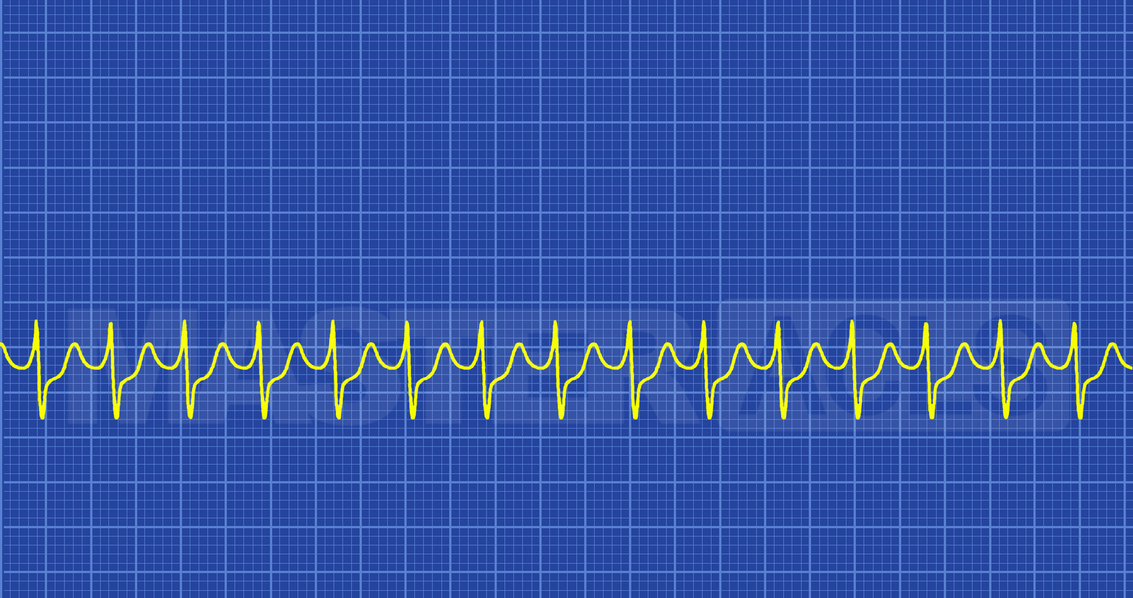
Technically Supraventricular Tachycardia (SVT) is a broad term that covers both atrial tachydysrhythmias (such as atrial flutter and atrial fibrillation) and atrioventricular tachydysrhythmias. However, in common practice most healthcare providers use the term Supraventricular Tachycardia or SVT to refer specifically to arrhythmias that are atrioventricular tachydysrhythmias. These arrhythmias arise from an abnormal electrical impulse produce by or dependent on the AV node.
Types of Atrioventricular tachydysrhythmias
- AV nodal re-entrant tachycardia (AVNRT) (most common form, >50%); involves a reentrant circuit in the AV node itself
- AV re-entrant tachycardia (AVRT); involves a reentrant circuit formed by aberrant myocardial accessory pathways between the atria and ventricles. Conduction may be orthodromic or antedromic down the normal His-Purkinje pathway, the latter tends to produce a wide QRS complex.
- Junctional ectopic tachycardia (rare)
- Non-paroxysmal junctional tachycardia (rare)
Causes of SVT
AVNRT; associated with:
- Myocardial ischemia
- Rheumatoid heart disease
- Mitral valve prolapse
- Medications, poisoning and toxic exposures (e.g. caffeine, theophylline)
- Pericarditis
- Dehydration
- Electrolyte Imbalances
AVRT; associated with:
- Presence of an accessory tract, often with evidence of a pre-excitation syndrome on the baseline ECG (e.g. Lown-Ganong-Levine syndrome)
- Ebstein’s anomaly
- Dehydration
- Electrolyte Imbalances
Junctional ectopic tachycardia and Non-paroxysmal junctional tachycardia
- Cardiac ischemia
- Digoxin toxicity
- Cardiac surgery
- Dehydration
- Electrolyte Imbalances
In all types of SVT symptoms result from the fact that the heart fills during diastole with diastole being approximately 2/3 of the cardiac pumping cycle and the faster the ventricular rate the less time the ventricles have to fill. If the ventricles don’t fill then the cardiac output drops causing less blood to be pumped to the extremities and less blood to be returned to the heard to feed oxygenated blood to the myocardium and be pumped again out to the body. This drop in cardiac output results in the following symptoms getting worse as the cardiac output continues to drop unless the ventricular rate is slowed.
Symptoms see in stable SVT patients
- SOB
- Fatigue
- Palpitations
- Numbness in Extremities
Symptoms see in Unstable SVT patients
- Chest Pain
- Hypotension
- Loss of Consciousness
Treatment
Consider and treat all underlying causes while providing symptomatic are and support for all stable patients. Vagal maneuvers and Adenosine may be used to slow stable WVT to make it a better perfusing rhythm or to convert stable SVT patients to a sinus rhythm. Unstable SVT should always be treated with synchronized cardioversion immediately. Recommended dose is 50-100 Joules. Drugs should not be used to treat unstable tachycardia.